Date: 16th December 2020
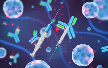
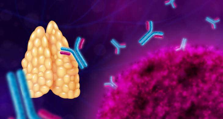
The thymus is a specialised primary lymphoid organ of the immune system, where T lymphocytes mature. A dysfunctional thymus can lead to severe immunodeficiency and there is currently an unmet clinical need for suitable thymic therapeutic strategies. Now, scientists have rebuilt a human thymus, using human stem cells and a bioengineered scaffold, which when transplanted into mice was able to support mature T cell development.
Attempts to rebuild a fully functional thymus have up until now met with limited success, likely due its inherent complexity. It is a bilobed organ with two subcomponents: the cortex and the medulla and is made up of epithelial, dendritic, mesenchymal and endothelial cells. However, clonogenic multipotent stem cells in the rodent thymus have been identified which were capable of contributing to the skin and thymic microenvironment after extensive ex vivo expansion.
Now, scientists from the Francis Crick Institute and University College London, UK, led by Yann Barrandon have identify epithelial-mesenchymal hybrid cells taken from patients who had to have the thymus removed during surgery, which were capable of long-term expansion in vitro, and were able to reconstitute an anatomic phenocopy of the native thymus. When transplanted into humanised immunodeficient mice, the bioengineered thymi were functional and able to support the development of mature and functional human T lymphocytes.
To start the team collected thmyi from 33 patients, ranging in age from 3 days to 11 years. The team then cultured dissociated cells from the donated tissue and identified clonogenic thymic epithelial cells (TEC) and thymic interstitial cells (TIC). Further investigations showed that these TEC co-expressed epithelial and mesenchymal markers but were distinct from the TIC and both were able to expand substantially under culture conditions.
Given the capacity to obtain billions of both sets of cells within a relatively short period of time the team then needed a 3D scaffold on which the cells could be seeded. The scaffold would act as a guide for cellular re-organisation creating a physiological pattern.
Here, they developed a unique microvascular surgical approach to obtain thymic extracellular matrix (ECM) by whole-organ decellularisation to create a natural thymic scaffold. In order to achieve this they cannulated the thymus of a rat, and used it to perfuse the organ with detergent and enzymatic solutions, so only the structural scaffold remained intact.
These scaffolds were then injected with up to six million human TEC and TIC, at an optimised ratio of 5:1, from the in vitro colonies the team had grown in the lab. The cells grew onto the scaffolds and after only five days, the organs had developed a similar stromal organisation to those seen in nine-week old foetuses. TIC were found to be crucial for re-organisation of TEC along the 3D ECM of the reconstituted scaffold.
Human thymus morphogenesis recapitulated in vivo
The team then wanted to see whether the repopulated thymus scaffolds would mature and sustain human T cell reconstitution from haematopoietic stem cells (HSC) in vivo. So they implanted the scaffolds subcutaneously into humanised severely immunodeficient mice, 4-6 weeks after they had been sub-lethally irradiated and then transplanted with human HSC. Promisingly, they found that in >75% of cases, the thymi were able to support the development of human lymphocytes.
Finally, the team wanted to determine whether the repopulated scaffolds could support peripheral T cell reconstitution where the engineered scaffolds were the only thymic stroma. Immune deficient mice, which were null for the thymus (Foxn1null) were implanted with repopulated or empty scaffolds. Upon bone marrow reconstitution no T cells were detected in the periphery of these mice when transplanted with empty scaffolds however, they were detectable in the mice treated with repopulated scaffolds.
Conclusions and future applications
The team here have demonstrated that the postnatal human thymus harbours TEC and TIC that can expand to clinically relevant numbers in vitro, enabling the reconstruction of a human functional thymus in vivo. The 3D natural ECM were shown to be crucial for thymus morphogenesis from these cultured cells, and the bioengineered thymi were able to attract circulating human HSC, support T cell development and repopulate the periphery in athymic mice. Interestingly, the human T cells that developed within the human scaffolds were functionally different in their response to stimulation from those which developed in the endogenous mouse thymus, demonstrating the instructive capacities of the human stroma.
The team are hoping to use the system to address many immunological questions. For example how different types of T cells develop and functionally mature and how the thymus helps the immune system to recognise self from non-self.
They are planning to develop the system further such that it can be used for medically relevant applications such as thymus transplants for conditions such as athymic DiGeorge syndrome. They are also looking into the possibility that the artificial thymus may be able to tackle host-versus-graft disease or alleviate transplant rejection. If a thymus was grown from cells taken from the organ donor, the new thymus could also be transplanted into patients receiving the donor organ, in order to prevent the organ being rejected.
Whilst, there has been ongoing efforts to bioprint transplantable organs in recent years, the ability to create complex 3D structures has been a hindrance in the field, such that bioprinting in space has even been explored. The team here circumvented this issue by developing a new technique which allows preservation of the fine 3D ECM meshwork of the thymus which created the scaffold. The team are now working on scaling up and refining the entire process with a view to building a human sized thymi, creating an artificial thymus which could be used in human transplants.
For more information please s the press release from the Francis Crick Institute
Campinoti, S., A. Gjinovci, R. Ragazzini, L. Zanieri, L. Ariza-McNaughton, M. Catucci, S. Boeing, J.-E. Park, J. C. Hutchinson, M. Muñoz-Ruiz, P. G. Manti, G. Vozza, C. E. Villa, D.-E. Phylactopoulos, C. Maurer, G. Testa, H. J. Stauss, S. A. Teichmann, N. J. Sebire, A. C. Hayday, D. Bonnet and P. Bonfanti (2020). “Reconstitution of a functional human thymus by postnatal stromal progenitor cells and natural whole-organ scaffolds.” Nature Communications 11(1): 6372.
https://doi.org/10.1038/s41467-020-20082-7