Date: 24th June 2021
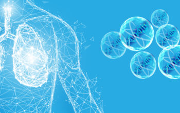
Like other respiratory illnesses, COVID-19 can cause lasting lung damage, and SARS-CoV-2 is capable of ravaging the respiratory and immune system by inducing proinflammatory cytokines. This causes lung complications such as alveolar oedema, hypoxaemia, dyspnoea and systemic inflammatory response syndrome and leads to severe illness. Whilst, global vaccination programmes are essential to fight the spread of the disease, progress has been varied, and there is still an urgent need to develop therapeutics for those who contract COVID-19 and suffer severe symptoms. Now, researchers have developed a cell mimicking nanodecoy, which acts a sponge to soak up SARS-CoV-2 thereby preventing it from entering lung cells and replicating, and which mitigates lung injury.
Angiotensin-converting enzyme 2 (ACE2) is present on many cell types and plays a pivotal role in host cell viral entry. SARS-CoV-2 has been shown to specifically attack ACE2-presenting type II pneumocytes in the lungs and goblet secretory cells in the nasal mucosa via its spike protein. As such, the spike protein in particular has become a popular therapeutic target. However, more aggressive variants associated with spike protein mutations are emerging, and therefore antiviral strategies focused on ACE2 on the host cells, which are not to subject to mutations, may provide a alternative and more efficient approach to developing new treatments.
Now, researchers at North Carolina State University and the University of North Carolina at Chapel Hill, US, led by Ke Cheng have shown that ACE2 nanodecoys derived from human lung spheroid cells (LSCs) can bind and neutralise SARS-CoV-2 and protect host lung cells from infection in vivo.
To start, the team converted individual LSCs into membrane nanovesicles, or nanodecoys, by serial extrusion through membrane filters with diminishing pore sizes, and showed these ‘bubbles’ expressed the ACE2 receptor and other lung cell-specific proteins on their surface. On average, one LSC generated 11,020 nanodecoys meaning large quantities of nanodecoys could be generated relatively easily.
Next, they tested whether the nanodecoys could bind to the SARS-CoV-2 spike protein via ACE2. Indeed, LSC-nanodecoys could recognise and competitively bind the spike S1 protein whilst other control decoys derived from cells expressing low levels of ACE2 did not. Furthermore, macrophages present in the culture had a higher efficiency to internalise the nanodecoys than the lung cells, suggesting this approach could rapidly clear the decoys and their neutralised SARS-CoV-2 by macrophages, and potentially other immune cells. The nanodecoys were functional in a dose-dependant manner, such that increasing the dose blocked more virus entry into the lung cells. Together, suggesting LSC-nanodecoys could protect host cells from infection via a SARS-CoV-2 mimics.
As the lungs are a primary target of infection, the team nebulised the nanodecoys enabling their administration via inhalation. The team found the decoys in the lungs of mice 72 h after a single inhalation treatment, and after 24 hours the majority were co-localised in macrophages.
Next, the team wanted to assess whether the inhaled LSC-nanodecoys could accelerate the clearance of a SARS-CoV-2 mimics in a mouse model. Mice were infect with the SARS-CoV-2 mimic and then 24 hours later were administered the therapeutic nanodecoys via inhalation. The amount of SARS-CoV-2 mimics were significantly reduced following inhalation of LSC-nanodecoys, a cytokine array analysis showed there was little proinflammatory cytokine induction, and the treatment appeared to be safe with no adverse effects.
To explore the nanotherapy further, the team turned to a more clinically relevant primate model. Cynomolgus macaques were challenged with SARS-CoV-2 by intranasal and intratracheal routes, then treated with the nanodecoy at days 2, 3, 4 and 5 post-challenge. High viral loads were recorded in the control animals indicative of replication, and in contrast the nanodecoy treated animals showed a dramatic reduction in viral load. This was accompanied by a significant reduction in the numbers of polymorphonuclear cells and neutrophils present in the lungs (alleviating inflammatory cell infiltration), and fibrosis was also decreased compared with the control cohort.
The team here have demonstrated that cell mimicking nanodecoys can reduce live SARS-CoV-2 infection and can protect lung cells from infection and damage. The nanodecoys were effective in alleviating inflammatory cell infiltration and decreased pulmonary fibrosis, and reduced viral loads in primates.
Whilst, the initial experiments show no adverse effects such as weight loss, fever, mortality, toxicity or histological changes this will have to be tested in more depth in order to translate this therapy for clinical trials. One area of focus will be how the nanodecoys are cleared from the body, but the primary investigations showed that the decoys were detected in the liver, kidneys and spleen of mice, suggesting clearance was via the reticuloendothelial system and are metabolised through the body, potentially via macrophages.
It is possible however, that translation into clinical trials may be accelerated as LSCs are already being tested in such studies. Furthermore, the ability to administer the therapy in a non-invasive manner, the fact that the nanodecoys are acellular, meaning they can be easily preserved and remain relatively stable longer term, will facilitate off-the-shelf use and clinical translation.
The idea of using ‘nanosponges’ or nanodecoys is not a totally new one. Recent studies using nanosponges constructed by wrapping polymeric nanoparticle (NP) cores with target cell membranes derived from lung epithelial cells and macrophages have also successfully been tested to protect against SARS-CoV-2 in mice. The work here, demonstrates the ability to administer and produce nanodecoys with ease and in large numbers, and will help lend weight to their use as a potential therapeutic agent for treating COVID-19.
For more information please see the press release from North Carolina State University
Li, Z., Wang, Z., Dinh, P.-U.C., Zhu, D., Popowski, K.D., Lutz, H., Hu, S., Lewis, M.G., Cook, A., Andersen, H., et al. (2021). Cell-mimicking nanodecoys neutralize SARS-CoV-2 and mitigate lung injury in a non-human primate model of COVID-19. Nature Nanotechnology.
https://doi.org/10.1038/s41565-021-00923-2