Date: 1st April 2021
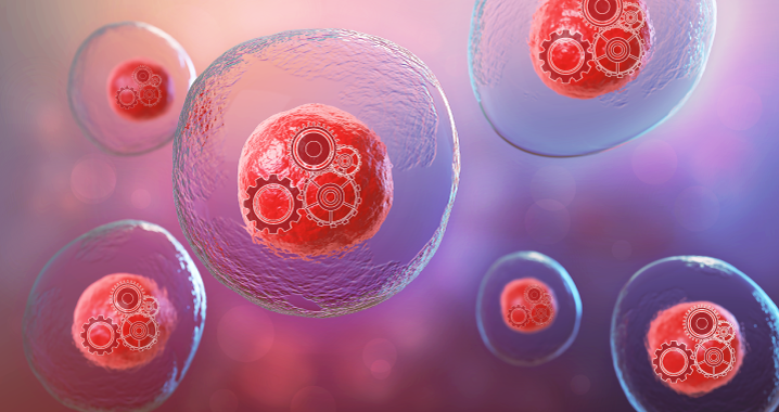
An artificial cell or genetically minimal cell is an engineered particle that mimics one or many functions of a biological cell. Such cells hold great potential, acting as biofactories to produce drugs, foods and fuels, detecting disease and producing drugs to treat them whilst living inside the body. Now, scientists create simple synthetic cells that grow and divide normally, helping us to understand the fundamental design rules of life.
Top-down strategies take a parent cell and manipulate it through genetic or protein engineering, with the resultant synthetic cell being closely related to its ancestor biologically. The construction of a top-down synthetic cells focuses on building using a minimal genome approach, where the cell’s DNA is pared down to those essential for life.
In 2010, scientists led by Craig Venter at the J. Craig Venter Institute (JCVI), US, constructed the first cell with an entirely synthetic genome, based on the bacterium Mycoplasma mycoides. They called it JCVI-syn1.0. Then 5 years later, JCVI-syn3.0 was ‘born’, containing the smallest genome of any-free living organism. Venter and his team, had reduced the 901 genes in JCVI-syn1.0 down to just 473 genes, with 149 of these essential genes classified with unknown or generic function.
However, whilst this was the simplest living cell ever known, an unexpected feature was JCVI-syn3.0’s cell divisions, which unlike the original synthetic cell and its parental wild type M. mycoides, produced wildly different shapes and sizes.
Now, researchers from JCVI, the National Institute of Standards and Technology (NIST) and the Massachusetts Institute of Technology, US, led by Elizabeth Strychalsk have identified 19 genes, including 7 needed for normal cell division that when added back to the minimal synthetic cell restore morphology and cell divisions, creating a new variant, JCVI-syn3A.
To determine the genetic requirements for normal cell divisions and morphology in the context of the minimal genome of JCVI-syn3.0, the team employed reverse genetics and microfluidic imaging of cellular growth. In order, to obtain time-lapse images the team designed a microfluid chemostat – which provided nutrients but shielded the cells from shear flow, which can affect the morphology of the mycoplasma.
Previously, during the process of genome minimisation from JCVI-syn1.0 to JCVI-syn3.0, genes or gene clusters were removed in various combinations from individual genomic segments. The team now identified that one of the strains called RGD6 (reduced genome design segment 6), showed a striking morphological similarity to JCVI-syn1.0.
The cluster contained 76 genes, the team then used a systematic, reverse genetics approach, to test the dependence of morphological variation first on gene clusters, and then on individual genes. One gene cluster containing 19 genes, showed ‘normal’ morphology similar to that of JCVI-syn1.0, and these genes restored the morphology of JCVI-syn3.0 when they were ectopically inserted into its genome. When the team tested these genes individually, 7 were found to be essential for morphology rescue, 2 had a known function in cell division, ftsZ and sepF, 1 was a hydrolase, and 4 were membrane-associated proteins of unknown function.
Conclusions and future applications
The team here present the first use of genomically minimised cells to determine the genetic requirements for the core physiological processes of cell division and the maintenance of cell morphology. They have created a new genetically minimal strain, JCVI-syn3A, which contained 19 additional genes to JCVI-syn3.0, and which restored more normal cell divisions and morphology. 7 of these genes were required and sufficient for this restoration, 5 of which had no known functions, adding new players to the field.
Their findings will have applications beyond mycoplasmas, and will inform bottom-up strategies to reconstitute cell divisions in synthetic cells. Furthermore, these strains will be an attractive minimal model for bacterial physiology and for broad engineering biology.
Understanding the minimal requirements for life will advance many biotherapeutics. Whilst ‘living’ medicines are being developed for the treatment of many diseases such as cancer and diabetes, new classes of artificial cells are also being created. Recently genetically controlled, stimuli-responsive artificial cells have been formulated to be functional under physiological conditions, synthesising therapeutic proteins on-demand in diseased tissues. The design and construction of a functional minimal genome is a key step to building cells and chassis for biotherapeutics, and this work reveals new genetic components required for cell division.
For more information please see the press release from NIST
Pelletier, J. F., L. Sun, K. S. Wise, N. Assad-Garcia, B. J. Karas, T. J. Deerinck, M. H. Ellisman, A. Mershin, N. Gershenfeld, R.-Y. Chuang, J. I. Glass and E. A. Strychalski “Genetic requirements for cell division in a genomically minimal cell.” Cell.
https://doi.org/10.1016/j.cell.2021.03.008


