Date: 14th January 2020
The current wave of next generation drug and therapeutic developments are being matched by the concurrent development of effective, safe, drug delivery systems. Now scientists have employed macrophages as vehicles for targeted delivery of drugs to the lungs.
Many therapeutic delivery systems rely on physical and biological differences between unhealthy tissue and surrounding healthy tissue in order to achieve targeted delivery. This model is however, somewhat simplistic and if you factor into this inherent cellular heterogeneity within the target tissue and nanocarrier surface manipulations needed to avoid eliciting a host immune response, complexity emerges and you encounter the challenges faced by the synthetic bioengineer in designing these delivery systems.
One area of emerging investigation is targeting whole organs rather than the cells or tissues. Some organs naturally sequester intravenously administered nanotherapies, such as the liver, spleen and lungs and these are potential go-to target organs for this approach. These organs also comprise the mononuclear phagocyte system (MPS), which is part of the immune system that consists of phagocytic cells, in particular macrophages, which can identify and isolate foreign materials and which also have a tendency to accumulate in the pulmonary system.
To harness this natural localisation to the lungs, therefore, scientists led by Ennio Tasciotti and Alessandro Parodi, from the Houston Methodist Research Institute, US, have developed a cell-based delivery system using macrophages as a delivery vehicle.
The work, published yesterday in Scientific Reports, was based on biomimetic exploitation of macrophages; a method previously pursued in the same lab for drug delivery purposes.
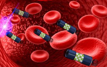
Cellular vectors or CELVECs, were generated using murine macrophage cells isolated from whole blood or grown using cell culture. Once purified, cells were loaded with a chemotherapeutic payload, DOX, using electroporation (DOX@CELVEC’s).
The loading of DOX, was shown to be directly proportional to the concentration of DOX in solution, and its release importantly displayed a delay in response when compared to passively-loaded cells. This was a crucial step in optimising timed-release of the payload to the lungs and not prematurely into circulation.
To initially assess DOX@CELVEC’s potential in efficiently inhibiting cancer cell growth, CELVEC cytotoxicity on human triple negative breast cancer cells was compared with free DOX treatments. An increase in cell toxicity was seen over and above that seen with free DOX, at day 1 and 2 post treatment, indicating further tests were worthwhile.
In order to determine the biodistribution of DOX@CELVEC, the team used intravital (live) microscopy and ex vivo organ analysis in non-tumour-bearing mice treated with DOX-loaded CELVECs. The lungs initially retained ~96% of injected cells with a 2.2-fold reduction occurring at 4 hours, and subsequent clearance of the system by the liver and spleen, as noted by an increase in cell accumulation in these organs.
Finally, the proof-of-concept experiment - a lung cancer mouse model. Here, mice showed an impressive 20-fold reduction in tumour signal following two weeks of treatment with DOX@CELVEC. Furthermore, a significant difference was observed between free DOX and DOX@CELVEC in as little as 4 days with significant levels of reduced inflammation in the lungs and the heart in mice treated with DOX@CELVEC over those treated with free Dox alone. With the apparent lack of acute side effects, the system showed high biocompatibility and represents a potentially new lung-targeted delivery system.
The conclusion here is that this system shows high reproducibility, saves time and is potentially more cost effective than current systems. The novelty of the method is that it also delivers a controlled release of payload, in this case with an optimal window of 4-6 hours, which corresponds to the load-bearing macrophages being resident in the lungs.
This system also adds to other advancements we have seen in developing delivery systems to the lungs, including the recent use of shuttle peptides to deliver CRISPRS safely and efficiently into the lungs via aerosols. As the lungs pose a particularly difficult organ to target and, with many debilitating diseases affecting this organ, expanding our repertoire of systems able to target the lungs is critical.
Whilst it is still early days for this method, it opens-up many new possibilities including the use of alternative cells lines and cargoes, and surface protein engineering to better target specific cell types in the lung. Also, as the CELVECs were observed in the spleen and then subsequently in the liver, by manipulating the timing of payload release it may also be possible to selectively target these organs in the treatment of metastases in more developed patients. Wherever this tech takes us we will be keeping an eye out for future developments.
Evangelopoulos, M., I. K. Yazdi, S. Acciardo, R. Palomba, F. Giordano, A. Pasto, M. Sushnitha, J. O. Martinez, N. Basu, A. Torres, S. Hmaidan, A. Parodi and E. Tasciotti (2020). “Biomimetic cellular vectors for enhancing drug delivery to the lungs.” Scientific Reports 10(1): 172.
https://doi.org/10.1038/s41598-019-55909-x