Date: 17th December 2021
Recent advances in high-throughput genomic sequencing have led to a detailed understanding of the genetic alterations that underlie human tumours. This has led to a ‘mutation-centric’ view of cancer which can drive precision medicine, with mutations being used as biomarkers to guide optimal therapy selection. However, there is mounting evidence that using mutations alone to determine therapies has its limitations and many cancer types still do not have available targeted therapies. It is also thought that the complexity of cancer extends to variations in cell transcriptional state however, the relationship between this and therapeutic sensitivity remains poorly understood. Now, researchers show that cancer cell state is plastic and has a dramatic impact on drug sensitivity. By altering the tumour microenvironment cancer cells can be driven from one state to another, opening new frontiers for drug development, selectivity and personalisation.
Pancreatic cancer has the highest mortality rate of all major cancers and its incidence is strongly related to age. Prognosis is often poor as there are no detections tools available for early diagnosis and patients often present late in the clinic. Despite extensive genomic characterisation of pancreatic ductal adenocarcinoma (PDAC), most cancers do not harbour therapeutically tractable alterations. However, bulk RNA-seq state measurements may mask heterogeneity at the single-cell level, and do not consider non-malignant cells which make up the tumour microenvironment (TME) - which plays a crucial role in tumour evasion and development. Therefore, understanding how cell state is shaped by the local TME and whether cell state can be used as a tractable biomarker for therapy selection remains of critical importance.
Now, researchers at Massachusetts Institute of Technology (MIT), US, led by Alex Shalek have shown that the TME drives cell state, plasticity, and drug response in pancreatic cancer, and prove that non-genetic modulation of cell state can strongly influence drug responses, uncovering state-specific vulnerabilities.
To start the team compared bulk and single-cell DNA and RNA-sequencing profiles of PDAC model systems (cell lines) and newly generated patient-derived organoids with primary and metastatic patient tumours to understand how well the model systems recapitulated patient RNA phenotypes. From the bulk experiments they found that model systems retained genetic fidelity well but lost expression state heterogeneity. Whereas primary and metastatic samples contained tumours with both classical-like and basal-like (aggressive) signatures, cell lines exhibited predominantly basal-like subtypes and organoids were nearly entirely classical-like.
However, single-cell RNA analysis revealed a third state, that appeared to be an intermediate between classical and basal, and co-expressed features of both programs to varying degrees, which they called intermediate co-expressor (IC) state.
Interestingly, when comparing each in vivo single-cell profile to the matched ex vivo organoid model, the researchers found that the organoids often existed in a different RNA state than cancer cells examined directly from the same patient. This suggested that the transcriptional state of the cells responded to changes in the microenvironment, and that growing them out of the body induced culture-specific programmes.
To unravel the role of the TME further the team tested whether specific aspects of the ex vivo culture environment governed cell state determination. Whilst the effects of extracellular matrix dimensionality were negligible, media composition did influence cell state. Here the found modulation of the media microenvironment allowed recovery of organoids to express more basal-like states which were previously lacking, and reciprocally cell lines could be induced to express more classical-like states. Together this suggested that culture medium-imprinting shaped the cell-state biases in extant model systems, and influenced their functional attributes.
Next, the team showed that transcriptional subtypes were associated with distinct immune microenvironments. Soluble factors specific to TME subtypes could drive cancer cell states and could influence their therapeutic responses. For example TGFB2 was the top differentially expressed factor shared by malignant cells in both basal and IC TMEs, treatment of organoids with TGF-β ligands shifted the classic state towards these two states. The duration of TGF-β exposure was concomitant with the degree of state shift, implying these states were highly plastic. Upon withdrawal of the signal a return to the classical-like expression was observed. Drug sensitivity also tracked with cancer cell state, for example organoids with the default classical state were sensitive to standard-of-care chemotherapeutic agents, upon TGF-β treatment for 3 weeks organoids were less sensitive to these agents, then the models were re-sensitised to the chemotherapy agents as TGF-β was withdrawn and they re-entered the classical state. Together, these data exemplified the remarkable phenotypic and functional plasticity inherent in tumour models.
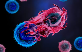
Finally, the team then used the organoid models for a drug screen, showing that different states were sensitive to different types of drugs, implying that the transcriptional state could be a major determinant of drug response. They also showed that paracrine signals from the local TME (originating from CD8+ T cells) such as interferons directed cancer cell transcriptional phenotypes and that similar effects were seen in patient biopsies where TME-supplied cytokines and immune activation are likely to play a critical role in directing cancer cell state.
Conclusion and future applications
The team here have presented a broadly applicable framework for aligning cancer cell states across in vivo and ex vivo setting, allowing them to identify drivers of transcriptional plasticity and the ability to manipulate this cell state to target associate vulnerabilities. They revealed a previously undiscovered relationship between TME and cancer cell transcriptional state, which can be used to drive drug selection.
The MIT team and colleagues are now performing much larger drug screens to measure how each drug affects pancreatic cancer cells in different states. Early experiments here already highlight site-specific differences in mesenchymal populations uncovering important differences between primary disease and metastasis. They will also extend their research to other types of cancer to determine whether different malignant cells exist in different states and can transition between states in response to microenvironmental changes.
The team envisage state-specific sensitivities of an individual’s cancer will become critical in selecting drugs and avoiding resistance. Treat a cancer in an unknown or incorrect transcriptional state with a particular drug it could target the wrong pathway and be resistant to treatment. By driving malignant cells into treatable states it could open new avenues for treating cancer.
This work should accelerate our understanding of the role of the TME which is increasingly becoming a new target for the development of new cancer treatments. Several of the most aggressive and deadly cancers that are resistant to immunotherapy are ‘cold’ due to immune exclusion. Recent works have identified two key players, DDR1 and cancer-associated mesenchymal stem cells, both of which play a role in immune exclusion, offering us new targets to improve tumour responses for resistant cancers. The ability to layer on to these types of research information about cell state will give us better understanding of the role the TME has to play, and give us the ability to hone into the interplay between drugs, cell state and the microenvironment. Ultimately it will lead to better treatments options for individuals and accelerate precision medicine.
For more information please see the press release at MIT News
Raghavan, S., Winter, P.S., Navia, A.W., Williams, H.L., DenAdel, A., Lowder, K.E., Galvez-Reyes, J., Kalekar, R.L., Mulugeta, N., Kapner, K.S., et al. (2021). Microenvironment drives cell state, plasticity, and drug response in pancreatic cancer. Cell 184, 6119-6137.e6126.
https://doi.org/10.1016/j.cell.2021.11.017