Date: 20th September 2021
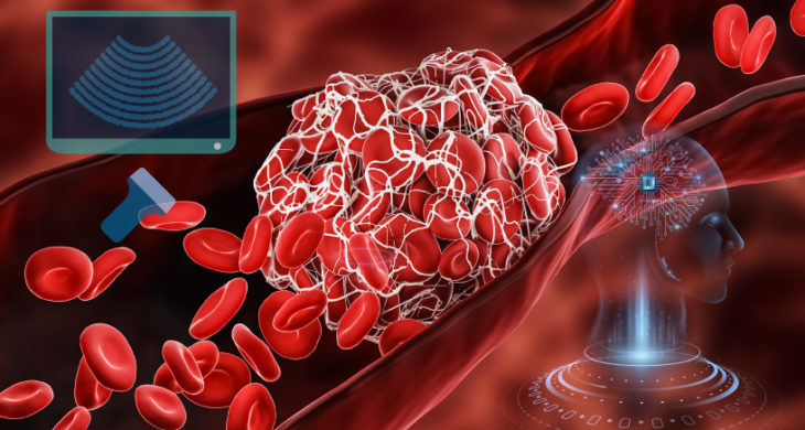
Deep vein thrombosis (DVT) occurs when a blood clot (thrombus) forms in one or more of the deep veins in the body, commonly in the legs. Early treatment is crucial, as clots can travel in the circulation and lodge in the lungs, known as pulmonary embolism (PE). Together, DVT and PE are known as venous thromboembolism (VTE) which is a leading cause of death and disability worldwide. Currently, there no reliable tests for DVT available that can be used in a general healthcare setting, and the diagnostic gold standard of compression ultrasound of the legs requires a specialist trained radiographer, resulting in long referral times and a large clinical burden. Now, a machine learning (ML) algorithm coupled to a hand-held ultrasound machine showed accurate diagnosis of DVT, even in the hands of non-specialist users, offering to cut waiting times, costs and unnecessary drug administration.
There are estimated to be ~10 million cases of VTE annually worldwide, and if left untreated DVT has a high level of morbidity with fatal clots moving to the lungs. Early diagnosis and treatment can often lead to recovery, but long-term complications and disabilities, such as post-thrombotic syndrome and chronic thromboembolic pulmonary hypertension, occur in 30-50% of patient who develop DVTs. However, many more patients present possible symptoms of DVT within the clinic, leading to long referral times for gold standard specialist-led diagnosis. This has serious consequences as many patient who do not receive diagnosis within 24 hours of suspected DVT are given unnecessary anticoagulants, with potential side-effects. Diagnosis at the point of care by non-experts is highly desirable, but has been an elusive goal to date.
Now, researchers at Oxford University, Imperial College and the University of Sheffield, UK, with study lead Nichola Curry, in collaboration with the technology company ThinkSono, based in London, UK, have demonstrated for the first time that ML artificial intelligence (AI) algorithms can potentially diagnose DVT, termed AutoDVT. AutoDVT coupled to a hand-held ultrasound machine and a smart device, provided guidance for free-hand ultrasound and aids non-specialists in detecting DVT.
The team started by training AutoDVT on preliminary data acquisition from 246 healthy volunteers, and 9 patients who were examined for DVT – 4 of which were positive. These acquisitions were performed by 2 radiologists and 3 trained engineers.
The team then used AutoDVT on internal and external validations sets. The external validation sets included 53 patients, 34 patients which were DVT positive and 19 DVT negative, confirmed by the current clinical pathway, from patients who presented to the Oxford Haemophilia and Thrombosis Centre, Oxford, UK, with symptoms suggestive of DVT. A further 30 patients with suspected DVT were recruited from the Clinic of Angiology, Ernst von Bergmann Klinikum, Potsdam, Germany - 4 of them were clinically confirmed DVT positive.
The algorithmic DVT diagnosis resulted in a sensitivity within a 95% CI (confidence interval) range of (0.82, 0.94), specificity of (0.70, 0.82), the positive predictive value (PPV) of (0.65, 0.89), and a negative predictive value (NPV) of (0.99, 1.00) when compared to the clinical gold standard, suggesting robust and accurate diagnosis by AutoDVT.
The team then wanted to test the system for operator skill level robustness, as the AI setup can guide non-expert users through the process. Directing the user using the ultrasound wand to the right locations along the femoral veins, enabling them to acquire the right images. 22 health volunteers were examined by 3 senior medical students, representing non-expert users. Each participant performed the examination twice in succession on the same given landmarks (guided by AutoDVT) and the average time for the second run was significantly faster than the first. Furthermore, >75% recorded the guided landmarks and compression sequences were reported as successful.
Conclusions and future applications
The work here provides a proof of concept that ML-based analysis can distinguish patients with and without DVT while providing image acquisition guidance for non-experts according to the clinical standard, and could potentially save health services $150 per examination.
It is the first study to show that machine learning AI algorithms can potentially diagnose DVT, and the researchers are due to start a test-accuracy blinded clinical study, comparing the accuracy of AutoDVT with standard care to determine the sensitivity of the for picking up DVT cases.
A NPV of around 99% is reassuring, meaning there is an extremely low chance that this prediction is wrong and that the patient might still have DVT. Whilst the PPV is robust, there is still a 20-30% chance of a wrong diagnosis meaning these patient might not have a DVT. However, a positive test with AutoDVT will always lead to a confirmatory scan with an expert, who can make treatment decisions which includes other criteria.
The emergence of AI in medical care is starting to offer new, accurate and powerful tools to advance a new generation of point-of-care applications. Here, AutoDVT will facilitate non-specialist healthcare professionals, such as GPs and nurses, to quickly diagnose and treat DVTs. Other AI-based systems such as early warning systems which flag potential sepsis patients and those that predict the ability to skip overnight vitals for patients all offer to improve patient care, and ease clinical pressure on struggling health-care professionals.
However, the diagnostic power of AI and ML is unprecedented, and now many applications are being developed such as predicting one-year all-cause mortality from echocardiograms, diagnosis of autism from maternal biomarkers, or early-stage breast cancer and COVID-19 associated pneumonia. However with AutoDVT, as with all of these applications, it is not likely that the clinician will become redundant, they are in the main aimed at accelerating work flows, aiding clinical decisions and reducing time to diagnosis. It is hoped they will ease the increasing burdens that healthcare systems are facing, providing better and more accurate treatments and improving patient outcomes.
For more information please see the press release at Oxford University or ThinkSono
Kainz, B., Heinrich, M.P., Makropoulos, A., Oppenheimer, J., Mandegaran, R., Sankar, S., Deane, C., Mischkewitz, S., Al-Noor, F., Rawdin, A.C., et al. (2021). Non-invasive diagnosis of deep vein thrombosis from ultrasound imaging with machine learning. npj Digital Medicine 4, 137.
https://doi.org/10.1038/s41746-021-00503-7