Date: 7th January 2021
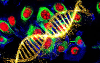
Progeria, or Hutchinson-Gilford progeria syndrome as it is also known, is a rare, progressive genetic disorder, which is lethal early in life, and sadly most children do not live past 13 years old. It is caused by a mutation in the LMNA gene and affects about 1 in every 4 million babies worldwide. In the majority of cases the disorder is attributed to a single-letter change in the DNA making it amenable to therapeutic gene editing. Now, scientists have used in vivo base editing to correct the single progeria mutation, rescuing the vascular pathology in progeria-modelled mice and extending the lifespan by nearly two and half times.
The LMNA gene encodes nuclear lamin A, a scaffold protein that provides structural support for the nucleus. The progeria mutation causes RNA mis-splicing which leads to a shorter mRNA transcript, which when translated, produces an abnormal protein called progeria. This toxic protein, induces rapid ageing as the abnormally shaped nuclear envelope causes defective cell division and DNA repair, and culminates in premature senescence and cell death.
Now, a team of scientists from the Broad Institute of MIT and Harvard, the National Institute of Health and Vanderbilt University Medical Center, US, led by David Liu, Jonathon Brown and Francis Collins have used an adenosine base editor to correct the pathogenic progeria mutation in patient fibroblast cells, and in mice harbouring the mutation. The in vivo base editing resulted in substantial correction of the mutation, rescued the vascular defects associated with progeria and greatly extended the lifespan of the mice.
The root cause of progeria is a single C-to-T DNA change, in order to correct this mutation the team used an ABE, which were first pioneered in the Liu lab. They started by testing the ABEs ability to correct the mutation in two primary fibroblast cell lines derived from progeria patients. They found up to 85% correction of the pathogenic mutation at 10 days post transduction, and up to 91% at 20 days, and this was accompanied by a significant decrease in mis-spliced LMNA mRNA (up to ~8 fold). Importantly, they found only a low frequency of bystander and off-target edits and found fewer abnormal nuclei in the treated cells.
Encouraged by these positive results the team wanted to test the system in a more clinically relevant model. They turned to assessing the ABE’s capability to directly editing the mutation in vivo in mice carrying the progeria mutation. These model mice exhibit many hallmarks of the disease, including progressive deterioration of the arterial system, skeletal changes, thinning fur, and premature death. Using an US Food and Drug Administration (FDA) approved adeno-associated virus (AAV) to deliver the editing machinery, the team administered cohorts of mice – 3 (P3) or 14 (P14) days after birth - with the therapy.
By 6 weeks of age the single injection of ABE-encoding AAV resulted in a modest to high level of correction (10–60%) of the causative LMNA point mutation, which was detected in various organs.
To assess the longevity of the treatment, the team also examined the mice at 6 months. This time they found notable differences between the P3- and P14-injected cohorts. Whilst, both sets showed an increase in DNA editing efficiency in several tissues compared to the six-week time point, the P14-injected group showed consistently higher levels of LMNA correction. In fact, on closer inspection the team found that progeria transcript decrease far out-weighted the DNA correction levels, suggesting that corrected cells may be more transcriptionally active than uncorrected cells or that cells with higher transcriptional activity may be more efficiently edited in vivo.
Next the team evaluated the progeria protein levels in various organs, key organs including the heart of animals had significantly lower levels of the proteins, in some cases a reduction of >80% was observed.
Importantly, this was accompanied by a histological rescue of the aorta. While the aorta of untreated progeria mice showed extensive loss of vascular smooth muscle cells and high levels of fibrotic cells accumulated around the aorta, the mice injected with the base editor appeared similar to samples from healthy mice.
Furthermore, lipodystrophy (reduced subcutaneous body fat) - which is a clinical feature of patients with progeria – was also modestly rescued in the treated mice.
Finally, the team wanted to determine any benefits the treatment may have on extending the lifespan of the animals. Over a 1.5 year period the mean survival of the P3-injected cohort was increased from 189 to 337 days in the control versus ABE- treated animals, whilst the P14-injected groups rose from 215 to an impressive 510 days for the ABE group – a 2.4 fold increase.
Of the mice that did not survive, analysis showed than a number of them developed liver tumours. This is a known long-term complication when using AAVs to deliver genes into mice, but thus far has not been reported in humans treated with therapeutic AAV vectors.
The study here is one of the first examples of using base editing in vivo to rescue a serious genetic disease. The team demonstrated that ABEs have high on-targeting editing (87-91%) with a low degree of off-targets effects.
In patients-derived cells, the ABE therapy could efficiently correct the pathogenic allele, substantially reduced RNA mis-splicing, decreased the abundance of progerin protein and rescued nuclear morphology abnormalities.
In progeria-model mice, ABE treatment resulted in durable correction of the pathogenic allele, amelioration of RNA mis-splicing, reduction of progerin protein in various tissues and greatly improved aortic health, almost fully restoring aortic pathology. Furthermore, this significantly extended the lifespan of the mice by nearly 2.5 fold, the team believe this would represent a ~5 to 6 year human equivalent.
However, whilst these data are undeniably very promising, adverse effects in the form of liver tumours were seen. Whilst, it is not clear whether a similar side effect would occur in humans, such results will guide future safety studies.
The team are now taking the results of the study forward into further preclinical studies, with the eventual goal of launching a clinical trial. Further optimisations such as using ABE variants with much higher editing activity than used here, or timing and dosing parameters will also be investigated.
The team are hoping that this work will provide a blueprint for the potential treatment of many other genetic diseases. It offers one of the first glimpses into the potential that base editing offers for in vivo gene editing.
Back in June last year, scientists used gene editing using a cytosine base editor (CBE) to repair a single nucleotide mutation in TMC1 mice in vivo, partially restoring hearing in mice. However, this made it possible to localise administration directly into the ear unlike in this study where the delivery route was systemic. These works, together with the recent demonstration that multiplexed precise base editing could edit up to three target sites simultaneously in primate embryos, places base editing as a strong emerging contender in the gene editing market and extends its range of applicable diseases and administration routes.
Whilst, new CRISPR gene editing clinical studies are rapidly emerging, most rely on ex-vivo editing. However, last year saw the first patient dosed with an in vivo treatment for Leber congenital amaurosis 10 (LCA10), an eye disorder, using CRISPR-Cas9. It is hoped that in vivo studies, such a seen here, will accelerate base editing therapies into the clinic, advancing patient outcomes and improving the quality of life for such patients.
For more information please see the press release from the Broad Institute
Koblan, L. W., M. R. Erdos, C. Wilson, W. A. Cabral, J. M. Levy, Z.-M. Xiong, U. L. Tavarez, L. M. Davison, Y. G. Gete, X. Mao, G. A. Newby, S. P. Doherty, N. Narisu, Q. Sheng, C. Krilow, C. Y. Lin, L. B. Gordon, K. Cao, F. S. Collins, J. D. Brown and D. R. Liu (2021). “In vivo base editing rescues Hutchinson–Gilford progeria syndrome in mice.” Nature.
https://doi.org/10.1038/s41586-020-03086-7